
In 2016, while I was completing my radiology training at the University of Pittsburgh Medical Center, AI pioneer Geoffrey Hinton famously suggested that medical schools should stop training radiologists. He believed computers would soon outperform physicians at interpreting medical images. [1] It was easy to see the logic. If AI could recognize patterns in pictures, medical imaging seemed like an obvious place for it to take over.
Ten years later, we’re facing the opposite problem.
Here in Pittsburgh, patients have reported waiting four to six weeks for imaging interpretations. UPMC and Allegheny Health Network have acknowledged staffing challenges, with UPMC citing a nationwide radiologist shortage. [2] A 2026 American College of Radiology analysis suggests shortages could persist for decades without meaningful changes to imaging demand or workforce capacity. [3]
Rather than becoming obsolete, radiologists have become increasingly difficult to find. My colleagues and I routinely receive three to five recruiting emails, texts, or calls per day.
So why hasn’t AI replaced us? And what’s taking so long?
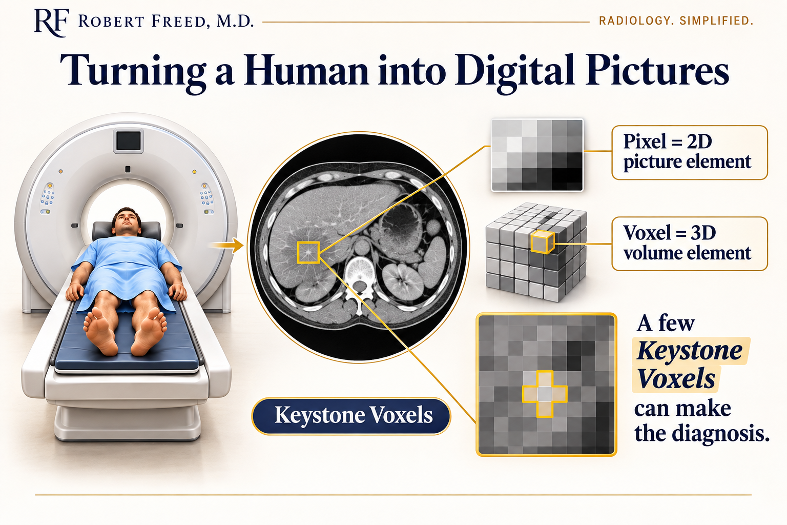
Turning a Human Into Pixels
Medical imaging turns information about the human body into digital pictures. For a CT scan, imagine dividing the body into millions of tiny boxes. A pixel is one of the dots on a screen. A voxel is its three-dimensional counterpart, with height, width, and depth. Each voxel represents a tiny piece of the body and is assigned a shade of gray.
We’ve reduced the extraordinary complexity of the human body to millions of little gray boxes.
AI can analyze all of them. The harder problem is knowing which ones matter.
Throughout my career interpreting chest, abdomen, and pelvis imaging, I’ve noticed that subtle diagnoses can hinge on a remarkably small number of critical voxels. As a proud Pennsylvanian, I call these “Keystone Voxels.” Their importance comes from their relationship to the anatomy around them. A tiny difference in location or shade of gray may provide the clue needed to recognize disease.
But those critical voxels aren’t always in the same place. Patients lie differently, organs move, anatomy varies, and disease evolves. I believe the early predictions underestimated both the variability of the human body and the unpredictable ways disease appears in individual patients.
When a Handful of Voxels Makes the Diagnosis
Consider appendicitis, one of the most common surgical emergencies and a frequent focus in appendicitis misdiagnosis and diagnostic delay cases. The appendix varies in size, shape, appearance, and location. It can sit behind the colon or extend deep into the pelvis. Inflammation can be subtle or severe, early or advanced, and patients have varying amounts of surrounding visceral fat.
Obvious appendicitis may be straightforward. But consider subtle appendicitis in an older patient whose anatomy reflects decades of aging, illness, surgery, trauma, or obesity.
The challenge is knowing which handful of gray boxes matter when disease is barely visible.
Why Medical Imaging Is So Difficult for AI
I believe three challenges explain much of AI’s difficulty with independent image interpretation.
1. The Patient: No Two Humans Are Identical
Identifying an abnormality depends on its contrast with the surrounding human body. The problem is that there is no single blueprint for what that background should look like. Genetics and development shape our anatomy before birth. Aging, obesity, disease, surgery, and injury add layers of variation over a lifetime.
One 75-year-old may look remarkably close to the textbook when you open the hood. Most will not. Their internal anatomy reflects decades of life.
Even the same patient is not identical from one scan to the next. Images obtained minutes apart can differ because of bowel peristalsis, cardiac motion, breathing, or movement during image acquisition. AI must recognize disease across an enormous range of what humans can normally, and abnormally, look like.
2. The Disease: No Two Cases Look Exactly Alike
Now introduce disease into those different bodies. Consider a bowel obstruction. The diagnosis may be the same, but its appearance can vary enormously. The obstruction can occur anywhere along the bowel. The degree of dilation varies. The bowel may contain mostly gas, mostly fluid, or both. The transition point may be obvious or subtle. Complications such as ischemia or perforation may or may not be present.
Diseases follow patterns, but those patterns aren’t absolute. AI must recognize thousands of diseases across countless variations of their appearance, superimposed on countless variations of human anatomy.
3. The Image: We Never See the Body Perfectly
Those tiny gray boxes don’t capture every detail. Some structures are smaller than the voxels themselves. Different tissues can appear nearly identical. Sometimes the difference between recognizing and missing disease is barely visible.
The patient changes the background. The disease changes the signal. The image determines how much of that signal we can see.
That’s a much harder problem than simply turning pictures into words.
Why More Training Data Isn’t the Entire Answer
Imagine teaching a child to recognize cows using photographs. You show them cows in grassy fields and other animals on city streets. The child gets every answer right. Then you show them a cow in a barn, and they don’t recognize it. Show them an empty pasture, and they say “cow.”
They learned the shortcut, not the animal.
AI can learn similar shortcuts, features that predict a diagnosis in training images but have little to do with the disease itself. A published study of AI detecting pneumonia on chest X-rays illustrated this problem. [4] More data can improve performance, but the examples must reflect the patients, diseases, and imaging conditions AI will encounter in practice. Otherwise, AI may pass the practice test while struggling with the next patient.
Some AI systems perform remarkably well at identifying a specific disease. But interpreting an entire examination requires looking for the unexpected, too.
Finding what you’re looking for is one challenge. Recognizing what you weren’t looking for is another.
What AI Developers Can Learn From Radiologists
Some of the most valuable information in an examination never appears in the radiology report. When I diagnose subtle appendicitis, a few sentences won’t capture the hundreds of images I examined, the structures I followed, or the tiny differences that caught my attention.
The report tells you what I found. It doesn’t necessarily tell you how I found it.
When AI misses a diagnosis, radiologists can help developers identify the Keystone Voxels and explain why they matter. AI may eventually discover patterns humans have never recognized. But understanding the clues human radiologists use could improve training, error investigation, and testing against difficult cases.
Teaching AI the diagnosis matters. Teaching it which clues make that diagnosis possible may be just as valuable.
When AI Is Confidently Wrong
Radiologists make mistakes. AI does too. What concerns me is how confident AI can appear when it’s wrong. If you’ve used ChatGPT on a subject you know well, you may have experienced this yourself. Without that underlying knowledge, mistakes are much harder to recognize.
Experienced radiologists remain essential for recognizing when an AI interpretation simply doesn’t fit.
Are We Training Our Replacements?
There’s an uncomfortable truth about radiologists helping develop AI: We’re effectively training our replacements.
Every subtle finding we identify and difficult case we explain brings AI closer to performing our jobs independently. I’m an actively practicing radiologist. These changes will reshape my own career.
But our responsibility is to put patients first. Patients are waiting weeks for imaging interpretations. Delayed diagnoses can mean delayed treatment, unnecessary suffering, and potentially preventable harm. My neighbor’s father waited five weeks for outpatient chest CT results. For families, these delays aren’t abstract workforce statistics. They’re real periods of uncertainty.
We have a nationwide radiologist shortage, growing imaging demand, [5] and technology that may help address both. Do we have an ethical obligation to help AI mature, even if that means training our eventual replacements?
I believe we do.
If AI can deliver faster, more accurate diagnoses, we should help develop it. I expect AI will eventually perform most medical image interpretation. The question is when.
For now, experienced radiologists working alongside AI offer the best path forward. But AI’s adolescence won’t last forever.
Will radiologists help shape what it becomes?
To learn more about my board-certified diagnostic radiology consulting or to discuss a complex imaging case, please contact me today.
