

Robert Freed, M.D.
Board-certified diagnostic radiology consulting
Radiology.
Simplified.
For trial counsel in medical malpractice, personal injury, and complex litigation — active full-time practice, objective interpretation, clear communication.
Academic Excellence
Reliable Educator
Real-World Volume
Peer-Checked
Robert Freed, M.D.
interpreted annually
Award, consecutive
radiologists in active peer practice
Radiology & Abdominal Imaging
Diseases of Expertise.

CT Chest
—
Pulmonary Embolism
Aortic Dissection
Lung Cancer
Pneumothorax
Pneumonia
Thoracic Trauma

CT Abdomen & Pelvis
—
Appendicitis
Bowel Ischemia
Bowel Obstruction
Bowel Perforation
Diverticulitis
Solid Organ Trauma

X-Ray / Ultrasound
—
Pulmonary Edema
Cholecystitis
Hydronephrosis
DVT
Ovarian Torsion
Testicular Torsion
Steps to Retention
-
01. Start a conversation
Reach out by text, email, phone, or through simple web-based form.
-
02. Retention
After our discussion to confirm subspecialty fit, you will receive several retention emails, including esignature digital contract, secure payment portal, and conflict check.
Visual Evidence
-

Demonstrative Exhibits
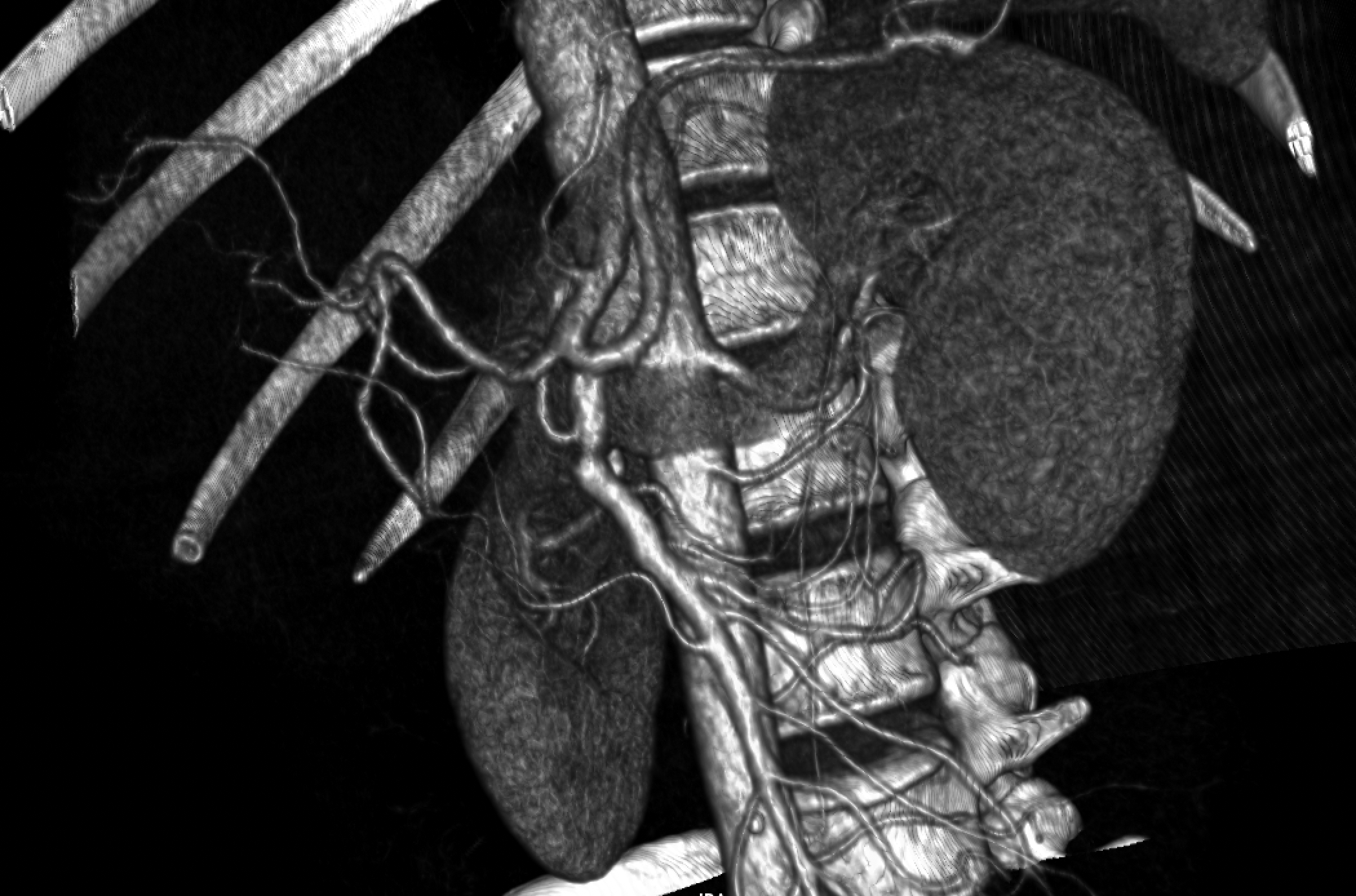
Annotated CT and MRI images, deposition slide decks, and side-by-side imaging comparisons, courtroom-ready.
-

3D-Printed Anatomical Models
Models Patient-specific anatomical models created from diagnostic imaging, allowing jurors to hold the anatomy in hand.
-

Quantitative Analysis
Volumetric measurements, interval change analysis, and objective imaging metrics that turn visual findings into measurable evidence.