
Demonstratives for Court
See the imaging.
Understand the medicine.
Dual fellowship-trained in Emergency Radiology & Abdominal Imaging Board-certified radiologist focused on the chest, abdomen, and pelvis.

-

Medical-Grade 3D-Printed Anatomy
Jurors can see and hold
-

Teaching Slide Decks
Complex imaging, explained step by step
-

Annotated CTs and MRIs
Highlighting the findings that matter most
-

Interactive 3D Renderings
Anatomy viewed from every angle
-

Quantitative Analysis
Measuring volume and size change over time
Turning medical imaging into something a jury can see, hold, and remember.
—
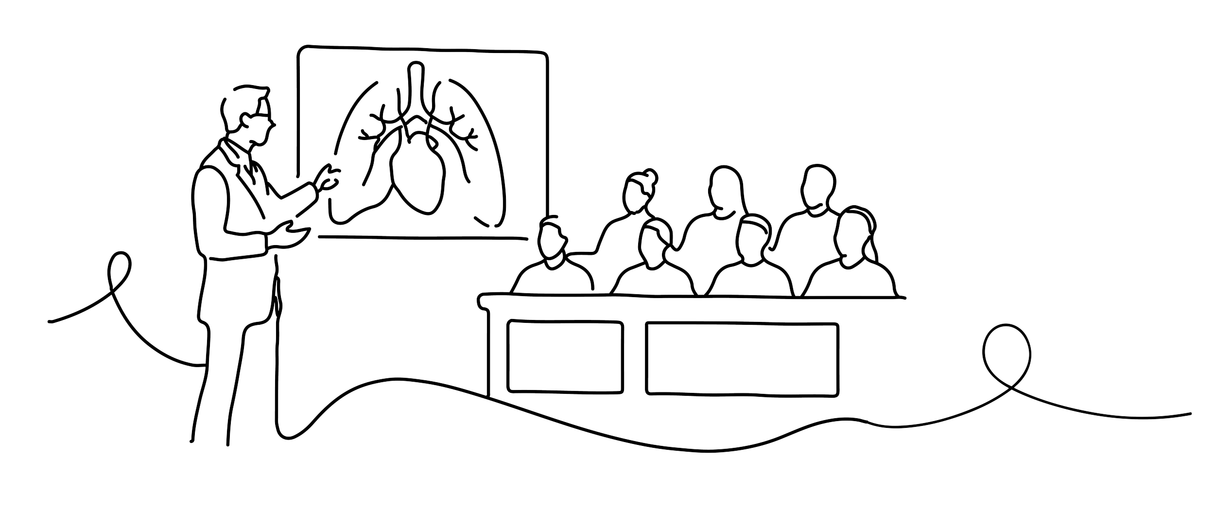
Medical imaging can be difficult to understand when you don’t look at it every day. I create clear, case-specific demonstratives that make important findings easier for attorneys and juries to see and understand.
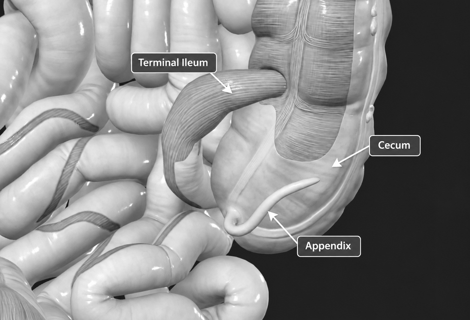
Demonstratives for Court
Exhibits translate complex radiologic findings into clear, compelling visuals that help attorneys and juries fully appreciate the anatomy and pathology at issue.
Jurors can hold a life sized model created directly from the patient’s CT scan.
Tumors, retained foreign bodies, vascular abnormalities, or blood clots that would otherwise exist only on a screen become real, tangible structures the jury can see and feel.