
The Missed Appendix: Diagnostic Delay and the Standard of Care
Appendicitis is one of the most common surgical emergencies and one of the top reasons a malpractice attorney will reach out to me for radiology consulting. For an attorney reviewing one of these cases, the central question may seem straightforward: Was appendicitis present on the imaging, and should the radiologist have diagnosed it?
The answer is often more complicated.
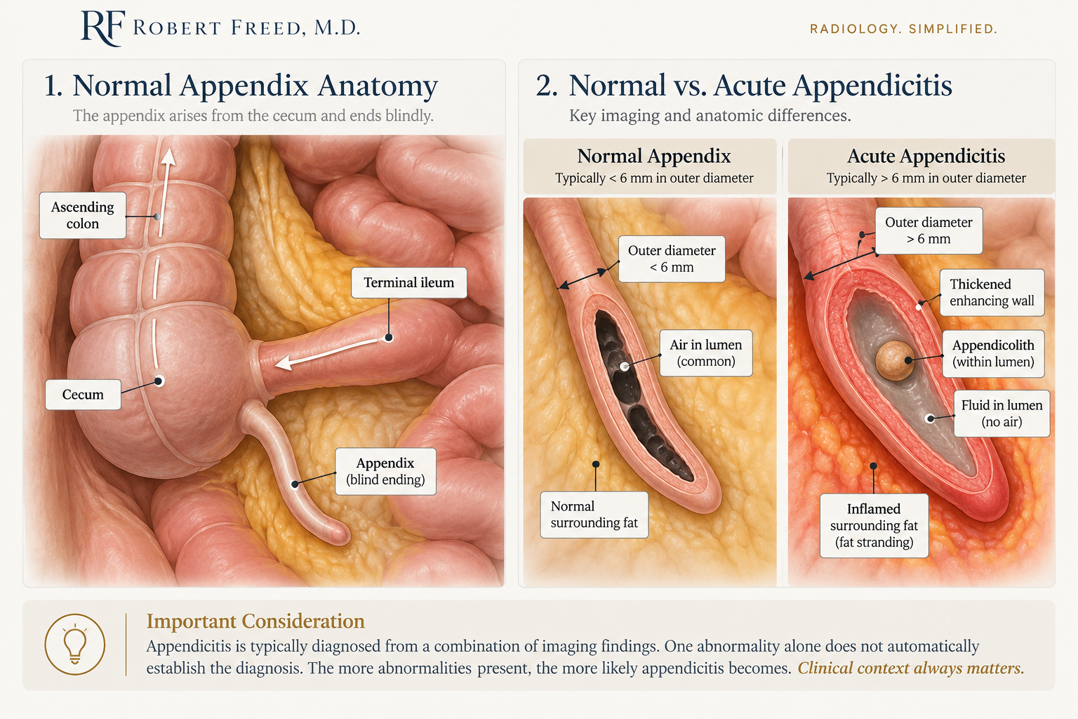
Finding the Appendix
The appendix is a small tube arising from the cecum in the right lower abdomen, near where the small bowel enters the colon. One feature is particularly important: the appendix ends blindly.
This matters on CT because the right lower abdomen contains multiple loops of small bowel that look like tubes when reviewed on individual imaging slices. The radiologist should follow the structure along its course and confirm that it ends blindly, think cul-de-sac, rather than continuing as small bowel, think main street.
Once the appendix is identified, another surprisingly useful question is: Is there air inside it? Air within the appendix is a strongly reassuring finding. Intraluminal gas is much more commonly seen in a normal appendix than in acute appendicitis.
A fluid-filled appendix deserves closer scrutiny. Fluid alone does not establish appendicitis, but the radiologist should look carefully for enlargement, inflammation in the surrounding fat, adjacent fluid, an appendicolith (a hardened stool ball within the appendix), wall thickening, and abnormal enhancement.
The clinical history then becomes particularly important.
Why Some Cases Are Harder Than Others
Appendicitis exists on a spectrum. Some CT examinations show an obviously enlarged, fluid-filled appendix surrounded by extensive inflammation. Early appendicitis may produce much more subtle findings.
Patient anatomy also matters. The appendix can extend into the pelvis, behind the cecum, or among adjacent bowel loops. Some patients have abundant abdominal fat separating these structures. Others have very little.
This is why having appendicitis and having obvious appendicitis on CT are not the same thing.
I think about diagnostic difficulty using three variables: how common the disease is, how typical the patient’s presentation is, and how obvious the imaging findings are.
Appendicitis is common. That does not mean every case is easy to diagnose.
What Did the Radiologist Actually Know?
Right lower abdominal pain, fever, nausea, and an elevated white blood cell count may raise suspicion for appendicitis. But there is another important question:
Were those findings actually conveyed to the radiologist?
A patient may have had three days of worsening right lower abdominal pain, fever, vomiting, and an elevated white blood cell count. But if the history provided to the radiologist simply says “pain,” much of that clinical context has been lost.
In a medicolegal review, the relevant information is not simply everything documented somewhere in the medical record. It is also what was reasonably available to the radiologist when the CT was interpreted.
A Missed Diagnosis Is Not Automatically Negligence
When appendicitis is diagnosed later, particularly after perforation or abscess formation, the earlier CT is often reviewed retrospectively.
Once the diagnosis is known, the reviewer knows exactly where to look and what abnormality to search for. The original radiologist did not have that advantage.
The standard-of-care question therefore is not simply:
Can we see appendicitis on the earlier CT now?
It is:
Should a reasonably prudent radiologist have recognized the findings at the time of the original interpretation?
That distinction is why I prefer to begin medicolegal cases with a blind review of the original imaging whenever possible. As a radiology expert witness, my role is to evaluate the imaging in the context available at the time rather than simply judging it with the benefit of hindsight.
Standard of Care and Causation Are Different Questions
Even when an earlier diagnosis should have been made, another question remains: What harm resulted from the delay?
Untreated appendicitis can progress to perforation and downstream complications including abscess, peritonitis, sepsis, and more complicated treatment. But the timing and consequences of that progression vary between patients.
Serial imaging can sometimes help reconstruct that timeline, demonstrating increasing inflammation, perforation, abscess formation, or other complications.
A proper review therefore separates several questions:
Was appendicitis present?
Was the appendix confidently identified?
How obvious were the findings?
What clinical information was provided to the radiologist?
Did the interpretation meet the standard of care?
Did any delay materially change the patient’s clinical course?
The final question often extends beyond radiology and may be better addressed by a surgical expert.
The presence of disease, diagnostic difficulty, standard of care, and causation are related questions. They are not the same question.
Understanding those differences is often where the real analysis of a missed appendicitis case begins.
To discuss a specific case or request a consultation, contact me today.
