
Radiology Expert Witness 3D Models: Bringing CT and MRI Findings Into the Courtroom
In many medical malpractice cases, CT scans and MRI studies are the most important pieces of evidence. However, the complexity of medical imaging can make it difficult for jurors and attorneys to fully understand the findings.
As a radiology expert witness, my role is to interpret the imaging, explain the abnormalities, and communicate those findings clearly in litigation. To make complex imaging easier to understand in court, I collaborate with Ricoh 3D for Healthcare, a globally recognized technology company with a strong presence in healthcare imaging solutions.

Through Ricoh’s healthcare division, attorneys now have access to a secure and highly sophisticated workflow that transforms medical imaging data into precise 3D printed medical models for trial and case preparation.
This platform includes:
• HIPAA compliant case portal for secure image transfer
• FDA 510(k) clearance medical imaging software used to convert CT (and occasionally MRI) scans into printable models
• Advanced medical grade 3D printers manufactured by Stratasys
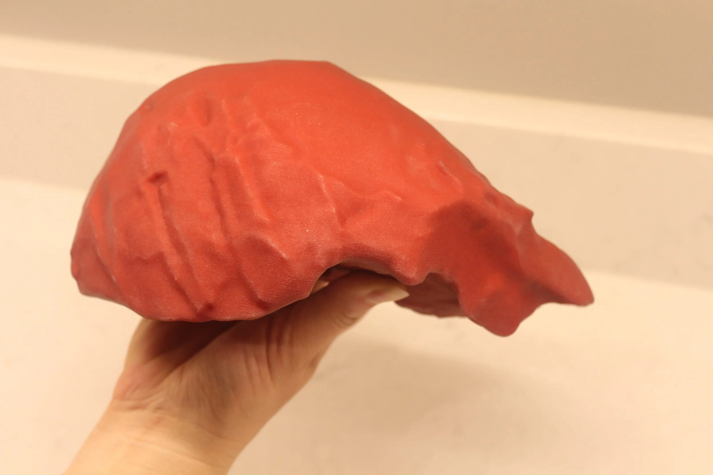
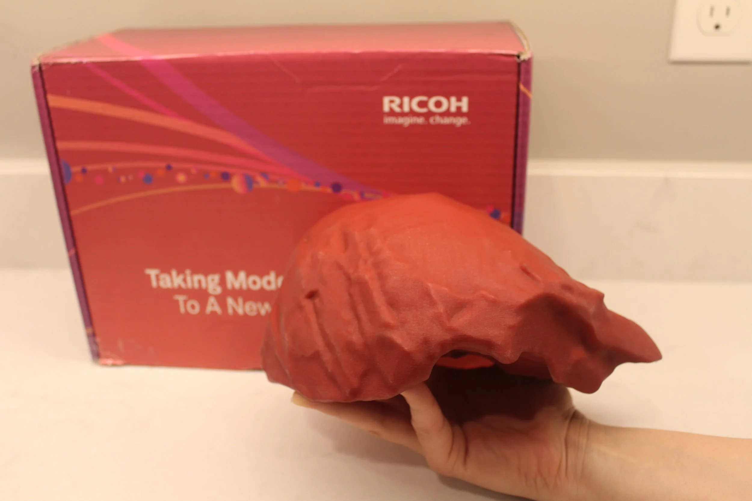
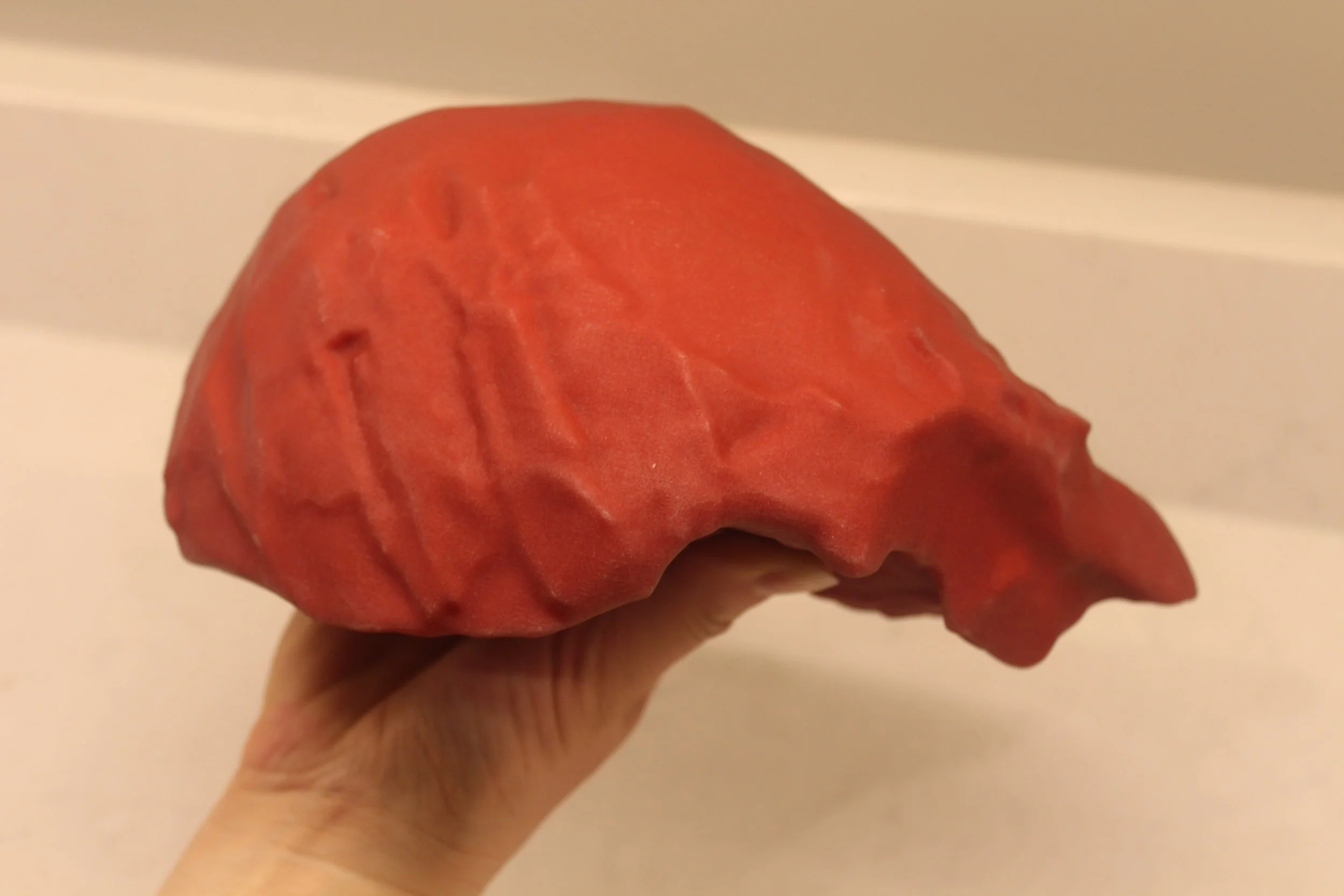
The result is a patient specific, life sized replica of the pathology identified on imaging.
How 3D Printed CT and MRI Models Are Created for Litigation
When a case involves a key imaging abnormality, I begin by carefully reviewing the patient’s CT or MRI study in my role as a medical imaging expert witness. The abnormality in question is precisely identified and segmented so the relevant anatomy can be accurately reconstructed. Ricoh’s medical engineering team then converts the imaging data into a high fidelity 3D printable model derived directly from the patient’s scan.
Attorneys retaining my services are able to provide input into several aspects of the final model, including:
• coloration of the pathology and surrounding structures
• filament type and material properties
• emphasis on anatomical relationships most relevant to the legal issues in the case
Once finalized, the model is produced using advanced medical 3D printing technology and shipped directly from Ricoh to counsel.
This process allows attorneys to present powerful demonstrative evidence based on the patient’s actual CT or MRI imaging data.
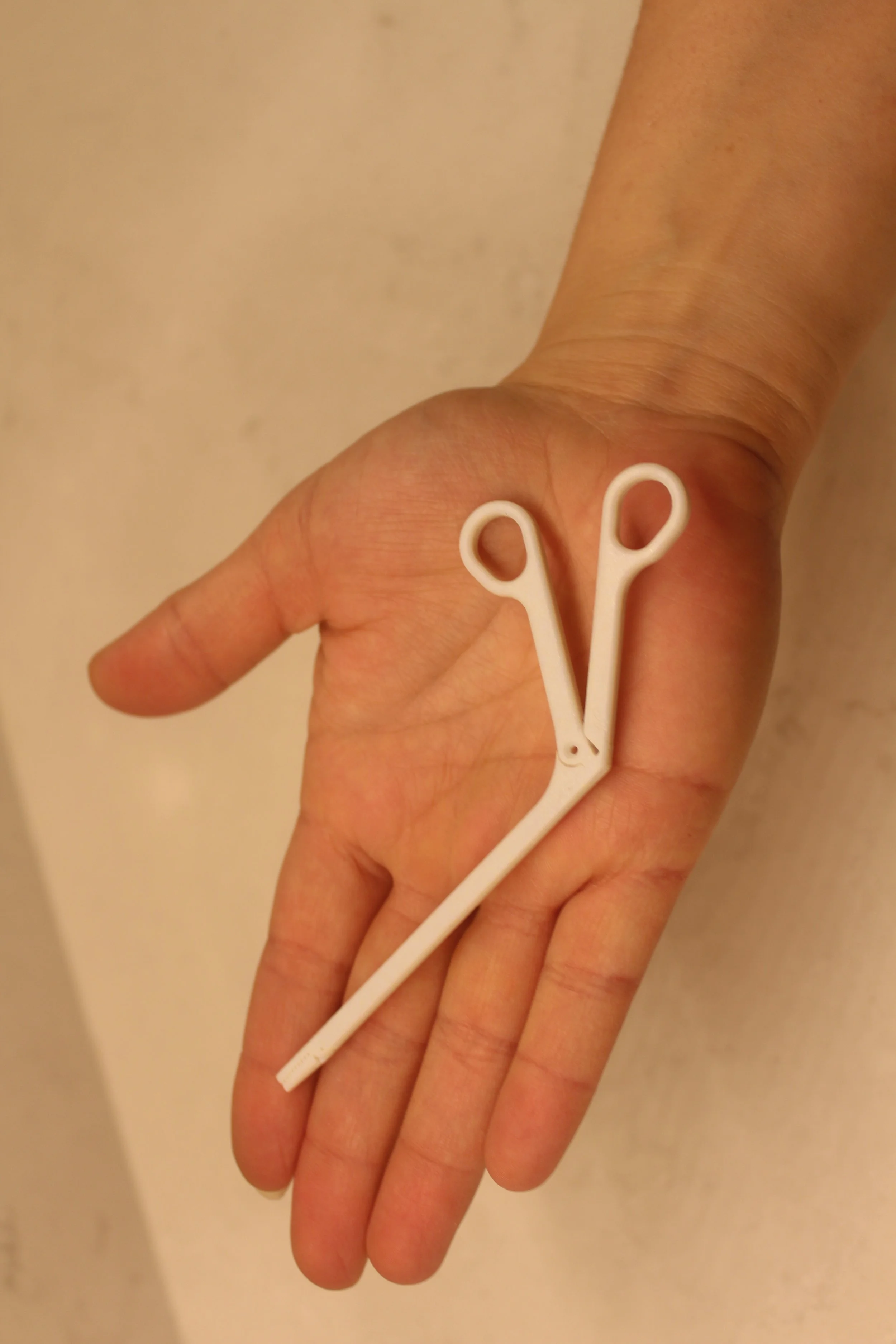
Case Example: Retained Surgical Foreign Body
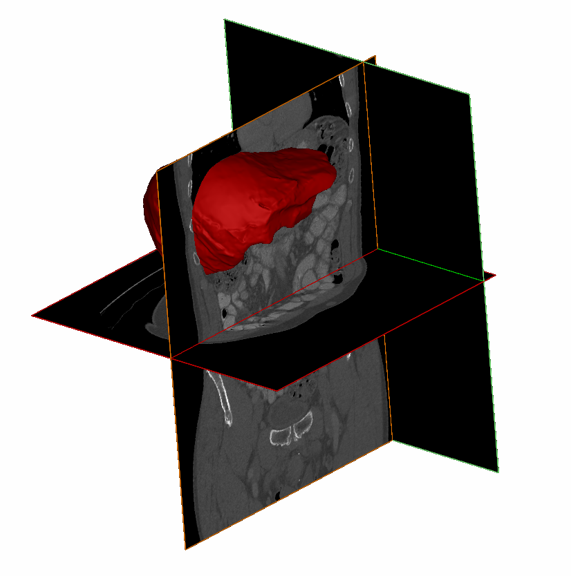
In a recent case involving a retained surgical sponge, the patient developed persistent abdominal pain following surgery. Multiple CT imaging demonstrated a retained object that had settled within the peritoneal space of the cavity.
Although the abnormality was visible on the CT scan, the spatial relationships between the foreign body and surrounding anatomy were difficult to explain using traditional radiology images alone. Using the patient’s CT imaging data, we created a life sized 3D printed model of the abdomen demonstrating the retained object and its precise location.
Once the model was reviewed during expert discussions, the anatomy became immediately clear. The case ultimately settled prior to trial, illustrating how physical models can dramatically improve understanding of complex radiologic findings.
Why 3D Printed Medical Models Are Powerful Demonstrative Evidence
Medical imaging is extraordinarily detailed, but for non physicians it can be difficult to interpret. A 3D printed CT or MRI model transforms abstract imaging findings into something jurors can see and hold in their hands.
Working with an established technology partner like Ricoh further reinforces the scientific rigor and professionalism behind the model’s creation. Jurors quickly recognize that the model is not a simple illustration, but a precise reconstruction derived directly from the patient’s imaging data.
When appropriate, these models can become some of the most powerful demonstrative exhibits in a medical malpractice case.